A reduction in mitochondrial activity has been strongly linked with various neurodegenerative diseases. Moreover, several mutations have been characterized that induce age-related mitochondrial malfunction. Scantox provides cell culture models that address the effect of compounds on mitochondrial activity and mitochondria related cell death in vitro.
Cell lines and primary neurons can be exposed to different toxins and examined for mitochondrial activity and mitochondrial membrane depolarization.
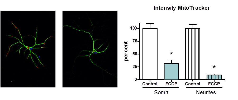
Primary E18 rat hippocampal neurons were cultured on poly-lysine coated glass coverslips until DIV8. Next, neurons were treated with FCCP, a protonophore and uncoupler of oxidative phosphorylation in mitochondria, for a defined period of time. Labeling of neurons with MitoTracker Red indicates active mitochondria. After fixation, neurons were labelled for MAP2. The effect of compounds on active mitochondria in soma versus neurites can be evaluated in individual neurons using Image Pro Plus software.

Figure: Effect of FCCP on active mitochondria in primary hippocampal rat neurons. FCCP treatment significantly reduced MitoTracker Red labeling in somata and neurites of primary rat hippocampal neurons (see graph). Images indicate (left) control neurons and (right) neurons upon FCCP treatment. MitoTracker labeling is indicated in red, MAP2 labeling in green.
Remark: This assay can also be performed with other mitochondria related toxins.
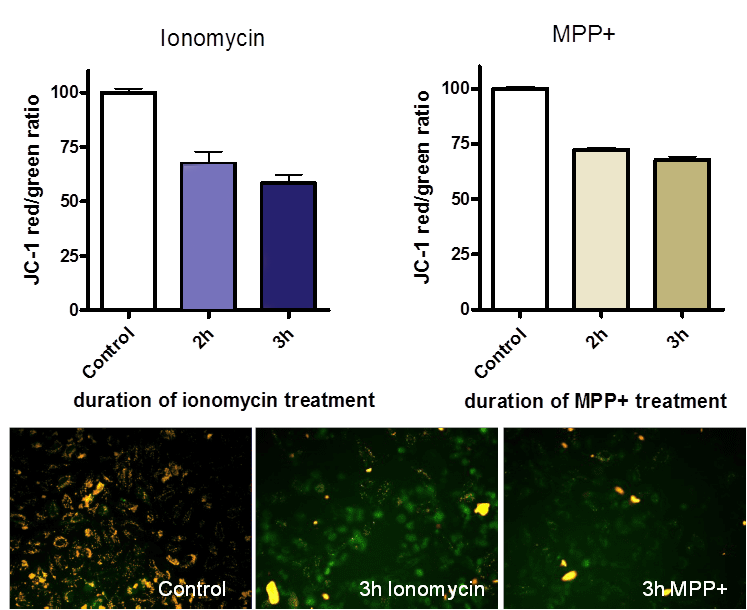
Differentiated SH-SY5Y human neuroblastoma cells were lesioned with the Calcium ionophore ionomycin or with MPP+. Lesions were examined with JC-1 for mitochondrial membrane depolarization and with the MTT assay for cell viability in 96-well plates. JC-1 labelling was determined in a 96-well fluorescence plate reader with the appropriate filter settings. The ratio of red/green fluorescence in control cells was set to 100%. Compounds can be examined for their ability to counteract mitochondrial membrane depolarization induced by selected lesion agents.

Figure: Effect of ionomycin and MPP+ on mitochondrial membrane depolarization by JC-1 labeling. Images indicate control cells and cells lesioned with ionomycin or MPP+ for 3 hours. In intact mitochondria, JC-1 forms red fluorescent aggregates, whereas green fluorescent monomers indicate mitochondria with depolarized membrane potential.
Remark: These assays can also be customized for primary neurons.
Effect of Mitochondrial Toxins and Ca2+ Influx on Mitochondrial Function
Compound testing for the following indications:
