A recent study from Niigata University researchers offers new insights into the protein-mediated motor neuron loss observed in amyotrophic lateral sclerosis (ALS). Using novel mouse models of ALS, the researchers focused on better understanding the role that the protein TDP-43 plays in the degeneration of motor neurons in ALS. Their findings, published in the journal Acta Neuropathologica, may help researchers better understand the mechanisms by which progressive neurodegeneration occurs in ALS, potentially leading to novel future therapeutic strategies for the disease.
Understanding ALS and TDP-43
Amyotrophic lateral sclerosis, or ALS, is a progressive neurodegenerative disease that affects the motor neurons in the brain and spinal cord. Over the course of the disease, the death of these neurons leads to widespread muscle weakness and paralysis; as ALS progresses, patients often lose the ability to speak, move, swallow, and breathe. There is no known cure for ALS, but when detected early, doctors can often slow the progression of the disease. Finding effective methods for early diagnosis has, therefore, been a primary area of focus for researchers.
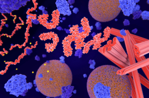
One marker associated with ALS is the accumulation of the TAR DNA binding protein (TARDBP or TDP-43). In 2022, a study published in JAMA Neurology found that TDP-43 accumulation in intramuscular nerve bundles may serve as an effective biomarker for early ALS diagnosis. But despite the known association between the abnormal accumulation of TDP-43 and ALS, this protein’s role in propagating degeneration in the disease has remained unclear.
That’s what a team of researchers from the Brain Research Institute at Niigata University sought to understand. As Dr. Osamu Onodero, a professor in Niigata University’s neurology department and this new study’s lead author, explains, “TDP-43 accumulation is seen in most of the patients with ALS, but there has been a long-standing debate on whether it propagates through the motor pathway and causes disease progression.”
TDP-43 Propagation in Mouse Models of ALS
To explore TDP-43’s role in ALS, Dr. Onodero and his research team developed ALS mouse models to accumulate TDP-43 in one of three specific areas: cortical motor neurons, spinal motor neurons, or skeletal muscles. By using these animal models, the team was able to observe how TDP-43 accumulation in each of those three specific areas would initiate the progression of the disease to other motor neurons.
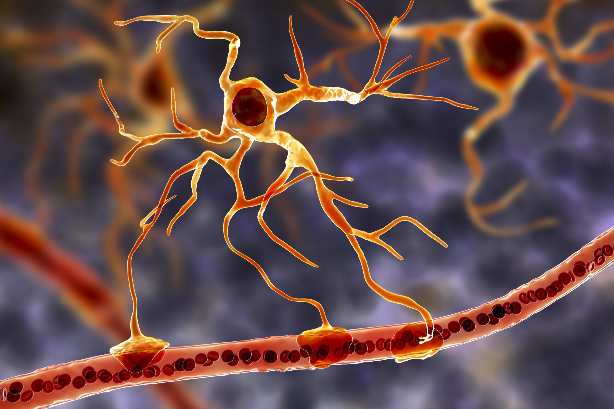
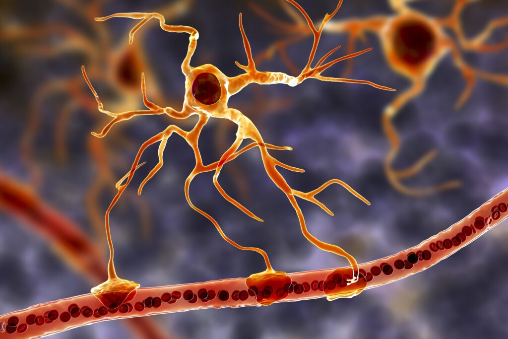
Dr. Onodero’s team observed that when TDP-43 was induced in cortical neurons, it caused mild degeneration. The protein was then “transported along the axons and transferred to the oligodendrocytes–non-neuronal cells that support neurons by wrapping axons with a protective layer called myelin to facilitate neuronal signal transmission.”
When TDP-43 was induced in spinal motor neurons, the results were drastically different: in these models, TDP-43 did not travel to nearby cortical or spinal neurons. In addition, unlike the mild degeneration that TDP-43 in the cortical neurons caused, TDP-43 accumulation in the spinal motor neurons triggered widespread and significant motor cell death, muscle atrophy, and motor dysfunction.
As the study’s co-senior author, Dr. Masaki Ueno, summarizes, “Our findings suggest that pathogenic TDP-43 has multiple properties to propagate degeneration in the motor pathways in ALS, probably by spreading itself and inducing other toxic events such as degeneration and inflammation.”
Implications and Future Research
The team’s findings suggest that in ALS, pathogenic TDP-43 has a variety of distinct mechanisms through which it can spread and propagate degeneration. Further research is needed to delve deeper into the mechanisms of how TDP-43 spreads and how it interacts with other proteins or cellular processes in ALS and other neurodegenerative diseases.
Still, these initial findings are significant. The more that is understood about the pathways that TDP-43 utilizes to trigger pathological events in ALS, the more informed researchers will be as they continue working to develop innovative therapies to prevent the progression of the disease.
Scantox is a part of Scantox, a GLP/GCP-compliant contract research organization (CRO) delivering the highest grade of Discovery, Regulatory Toxicology and CMC/Analytical services since 1977. Scantox focuses on preclinical studies related to central nervous system (CNS) diseases, rare diseases, and mental disorders. With highly predictive disease models available on site and unparalleled preclinical experience, Scantox can handle most CNS drug development needs for biopharmaceutical companies of all sizes. For more information about Scantox, visit www.scantox.com.