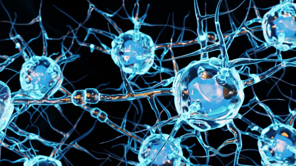
Swedish researchers have created the most detailed cell atlases of the human brain available to date. In two parallel projects, the research team from Karolinska Institutet analyzed millions of cell nuclei from human brains across a range of developmental stages. Chief among the study’s wide-ranging findings was the broad diversity of neurons found in the brainstem. The study’s authors hope their findings will help researchers better understand how the brain develops and is organized over time, laying the groundwork for future medical advances.

Exploring the Diversity of Brainstem Neurons and More Brain-Cell Census Findings
An advanced understanding of the composition and development of a healthy human brain is critical to treating neurological diseases as they arise. But while researchers have created comprehensive atlases of the mouse brain, until recently, there has been no such equivalent for the human brain. Indeed, outside of the cerebral cortex, few regions of the human brain have been thoroughly profiled.
That is changing thanks to two parallel studies conducted by researchers from Karolinska Institutet in Sweden and recently published in Science. As Sten Linnarsson, professor of molecular system biology in Karolinska Institutet’s Department of Medical Biochemistry and Biophysics, puts it, “We’ve created the most detailed cell atlases of the adult human brain and of brain development during the first months of pregnancy.”
In other words, as Linnarsson sums up, “You could say that we’ve taken a kind of brain-cell census.”
RNA Sequencing
Linnarsson’s group member Kimberly Siletti led the team’s first study, working in partnership with Ed Lein at Seattle’s Allen Institute for Brain Science. Together, the researchers used RNA sequencing to analyze the genetic identity of individual cell nuclei in three donated human brains.
Their analysis spanned more than one hundred distinct brain regions, including the forebrain (cerebral cortex, hippocampus, cerebral nuclei, hypothalamus, and thalamus), the midbrain, and the hindbrain (pons, medulla, and cerebellum).
The team identified more than 3,000 distinct cell types across these regions. Roughly 80 percent of these cells are neurons, which play essential roles in brain functions ranging from cognition and memory to sensory perception, movement, and emotion.
The team broadly found that neurons vary widely across brain regions and that “each brain area contains a specific complement of cell types and states.” However, within regions, subtypes of neurons did not follow clear distribution rules.
Where the greatest diversity of neurons were found was equally of note.
“A lot of research has focused on the cerebral cortex,” Professor Linnarsson notes, “but the greatest diversity of neurons we found in the brainstem. We think that some of these cells control innate behaviors, such as pain reflexes, fear, aggression, and sexuality.”
Cells Across Embryonic Development Stages
The billions of neurons and glia comprising the human brain across regions have distinct developmental origins. The hundreds of brain regions, and the many distinct cell types they contain, are established during an embryo’s first trimester of development.
Here was the basis, then, for the team’s second project, led by Emelie Braun and Miri Danan-Gotthold in collaboration with Sweden’s Human Developmental Cell Atlas consortium. Their project focused on analyzing and mapping over a million individual cell nuclei from embryos between 5 and 14 weeks of fertilization. The comprehensive atlas of embryonic brain development during the first trimester provides researchers with a clear picture of how the human brain forms over time. It creates a map of the spatial, temporal, and transcriptional changes that occur in the brain during this period.
These brain atlases will be freely available to researchers around the world. Ideally, this will provide a useful comparison tool for researchers studying neurological diseases, potentially helping them to identify more effective therapeutic targets over time.
Scantox is a part of Scantox, a GLP/GCP-compliant contract research organization (CRO) delivering the highest grade of Discovery, Regulatory Toxicology and CMC/Analytical services since 1977. Scantox focuses on preclinical studies related to central nervous system (CNS) diseases, rare diseases, and mental disorders. With highly predictive disease models available on site and unparalleled preclinical experience, Scantox can handle most CNS drug development needs for biopharmaceutical companies of all sizes. For more information about Scantox, visit www.scantox.com.


