Many of the motor and non-motor symptoms that occur in Parkinson’s disease are caused by the loss of midbrain dopaminergic neurons. Cell therapy has shown promise as a potential treatment for Parkinson’s disease by replacing these lost dopamine neurons, but successful cell therapy development has historically been hindered by poor graft survival. However, a recently published study from researchers at Mass General Brigham highlights a major breakthrough in successfully utilizing regulatory T-cells to enhance neuronal cell therapy outcomes in rodent models, demonstrating the potential of using personalized cell therapies for Parkinson’s disease treatment.
Parkinson’s Disease Treatment Limitations
Parkinson’s disease is one of the most prevalent neurodegenerative disorders worldwide, second only to Alzheimer’s disease in the United States. The current standard of care for Parkinson’s disease primarily involves dopamine replacement therapy, which has notable limitations. It is only able to target some symptoms of tremors and stiffness, and it comes with serious side effects, including dyskinesia, hallucinations or delusions, and compulsive behaviors.
Cell Therapy: Challenges and Breakthroughs
An appealing potential treatment alternative for Parkinson’s disease is cell therapy – specifically, cell therapy aimed at replacing the midbrain dopamine neurons that are lost in Parkinson’s disease.
Clinical trials using cell replacement therapy for Parkinson’s disease began in the 1980s. However, these trials have historically been hindered by one major issue: poor graft survival, with only 3-20% of grafted dopamine neurons surviving the procedure. Over the years, researchers have attempted various modifications to improve cell survival in transplantation. One of the first major advancements occurred three years ago, when personalized cell therapy demonstrated success in replacing dopamine neurons in a sporadic Parkinson’s disease patient. But even then, the limited graft survival remained a challenge.
New Treatment Breakthrough
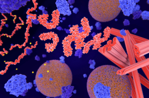
In a new study, Dr. Kwang-Soo Kim and his team at Mass General Brigham approached this problem with a novel hypothesis: that poor survival of grafted neurons was caused by “needle trauma” triggered by the surgical procedure itself and that co-transplanting regulatory T-cells with the dopamine neurons could help maintain immune homeostasis.
To test their hypothesis, the research team transplanted dopamine neurons into 6-OHDA rodent models of Parkinson’s disease. They were able to observe that the surgical procedure triggered “needle trauma,” which encompassed both acute inflammation and an adverse immune response in the brain tissue itself.
In the next stage, the team co-transplanted regulatory T-cells along with the dopamine neurons. The results were significant: more of the grafted neurons survived, and behavior recovery was “faster and more robust.” Dr. Kim and his team further observed that the co-transplanted T-cells were able to significantly suppress the outgrowth of reactive inflammatory cells in the brains of the 6-OHDA lesioned rodent models.
As Dr. Kim explains, “This finding is very significant because a potential hazard associated with cell transplantation is often the outgrowth of undesirable, potentially harmful cells. The most important criterion for cell therapy is safety.”
Implications for Neurodegenerative Disorders
The benefits of using regulatory T-cells to mitigate needle trauma and improve cell survival are not limited to Parkinson’s disease. Dr. Bob Carter, a co-author of the study and Chief of Neurosurgery at Mass General Hospital, noted that these same principles can be applied to other neurodegenerative disorders, such as Alzheimer’s, ALS, or Huntington’s disease.
Improving the delivery, survival, and recovery of neuronal cell therapies could represent a significant breakthrough in harnessing the therapeutic potential of personalized, stem-cell-based therapies to treat Parkinson’s disease and other neurodegenerative conditions.
While this study demonstrated promising results in rodent models of Parkinson’s disease, there remains a need to further investigate and understand the precise mechanisms by which regulatory T-cells enhance the survival of dopaminergic neurons.
Scantox is a part of Scantox, a GLP/GCP-compliant contract research organization (CRO) delivering the highest grade of Discovery, Regulatory Toxicology and CMC/Analytical services since 1977. Scantox focuses on preclinical studies related to central nervous system (CNS) diseases, rare diseases, and mental disorders. With highly predictive disease models available on site and unparalleled preclinical experience, Scantox can handle most CNS drug development needs for biopharmaceutical companies of all sizes. For more information about Scantox, visit www.scantox.com.